The Great Brain Debate: Nature or Nurture? (2004)
Chapter: 5 Controversies: New Neurons and Genes and Behavior
5
CONTROVERSIES: NEW NEURONS AND GENES AND BEHAVIOR
The previous chapter described various ways cortical circuitry can be modified—from simply altering the strength of synapses to neurons extending new branches and making new synapses. It deftly avoided one of the most contentious questions of the day: Can new neurons be generated in the adult mammalian brain? New neurons can be generated in nonmammalian brains, as I shall describe below, but what about the mammalian brain? This is a hotly debated issue that has implications not only for understanding the adult mammalian brain, but perhaps even more so for the aging brain. It has implications too for the extent of plasticity that can occur in the mammalian brain, also a topic not touched upon in the last chapter but one of vital importance.
The second half of the chapter considers another very contentious issue—the relation of genes and behavior. This is a topic about which reams have been written and which generates enormous heat because of its obvious social implications and the high stakes involved. If our behavior is fixed by our genes, why should
we try to alter it by education or social programs? Obviously, in half a chapter I can only touch on the topic, but hard neurobiological facts are few and far between here. This discussion, then, can only point the way; its resolution—if indeed it even can be resolved in a satisfactory way—is not yet clear.
Generating New Neurons
Neurons differ from other cells in the body in at least two important ways. First, neurons have an absolute requirement for oxygen. Deprived of oxygen, mammalian neurons die in just a few minutes. Other cells can survive for some time without oxygen and even function for a while by a fermentation-like process that breaks down sugar (glucose) in the cells to smaller molecules, thereby producing the energy needed for them to keep functioning. During a 100-yard race, a runner’s leg muscles probably use up the available oxygen in the first 30 yards or so. The rest of the race is run without oxygen (anaerobically). After the race, the muscle cells break down the smaller molecules using newly available oxygen. While this is going on, a runner typically breathes hard to repay the oxygen debt built up in the muscles during the race. After strenuous activity, muscles often feel sore, due in part to the buildup of the breakdown products of glucose, particularly lactic acid, during the anaerobic phase of muscle activity.
Neurons, on the other hand, cannot survive anaerobically; they stop functioning shortly after the oxygen runs out. After a heart attack or stroke in which blood flow to the entire brain or part of the brain is shut off, neurons begin to die after just four to five minutes. Other tissues can survive much longer, so that if oxygen is eventually restored, they survive, but the brain does not. Thus today, with our sophisticated life support systems, someone suffering a heart attack might be left permanently brain dead or with permanent brain damage even though other organs such as the heart, liver, and kidney recover completely.
The brain’s requirement for oxygen is so acute that when a part of the brain is active, blood flow to that region rapidly in-
creases. The rise in blood flow to an active part of the brain can be measured and is the basis for certain imaging techniques, including positron emission tomography (PET) scanning and functional magnetic resonance imaging (fMRI), that enable neuroscientists to observe which parts of the brain are especially active when a subject is carrying out a task. These noninvasive imaging techniques are adding enormously to our understanding of human brain function.
The other way neurons differ from other cells is that once they have differentiated from a neural progenitor cell into a specific type of nerve cell, they never divide again. As I pointed out in Chapter 1, the brain initially overproduces neurons, so that during the first year of life humans have as many neurons as they ever will. Thereafter, the number of neurons decreases throughout life. A controversial issue is how much cell death normally occurs throughout the life span—a topic that will be taken up in the next chapter.
Because neurons can’t divide again after differentiation, and because anatomists observed that after injury to various parts of the brain no new neurons seem to appear, it has generally been believed that new neurons are not generated in the adult mammalian brain. This is in marked contrast to other parts of the body where, after injury, new cells are rapidly generated and the injury is repaired. A cut on the skin heals quickly as new cells are generated and fill in the defect. Usually within a few days no sign of the cut can be detected. “New glial cells are generated in the mammalian brain throughout life, but not neurons” was the dogma.
Some recovery, of course, is usually observed following brain injury or stroke, but this is thought to be the result of the recovery of cells damaged but not killed by the lack of oxygen, or the remaining brain cells taking over for the lost cells. For example, as has been clearly shown and was discussed in the last chapter, after brain injury, neurons sprout new branches and form new synapses, allowing for at least some recovery. The fact that neuronal brain tumors are not seen in adults is another piece of evidence supporting the notion that new neurons are not generated
in the adult brain. Brain tumors in adults are mainly glial cell tumors or tumors arising from other nonneuronal cells. In children, rare neuronal cell tumors are observed, mostly derived from a pool of neuronal precursor cells that continue to proliferate for a time after birth.
A New View
The dogma that the mammalian brain cannot generate new neurons was recently challenged. The challenge arose, not from disputing the notion that neurons, once differentiated, no longer can divide, but from the discovery that two regions of the adult mammalian brain, the hippocampus and the cerebral subventricular zone (SVZ), retain neural stem cells that generate new neurons. These new data are convincing and fly in the face of the old dogma. But what are these new neurons up to? What role do they play? As I shall describe below, these questions are yet to be answered.
The finding that two brain regions can generate new neurons has spurred investigators to examine other brain areas, including the cerebral cortex, for similar neural progenitor cells. Claims that such cells exist in the cortex have been made with much fanfare, but these findings are hotly disputed. Glial cells continue to divide in the adult cortex and one view is that the dividing cortical cells are glial progenitor cells. Further confusing the picture is the view of some researchers that glial progenitor cells can and do become neurons. Others disagree, believing that new neurons are generated in the cortex, so the story is murky at the moment. Most neuroscientists are quite agnostic on the issue of whether most regions of the brain are capable of generating new neurons.
More significant, perhaps, than whether there are new neurons produced in the mammalian brain is the question of the role they play: What is their significance? Again, here the story is incomplete, but also strange. We know most about the hippocampal cells, which Fred Gage and his colleagues at the Salk Institute in La Jolla, California, have studied extensively. Less is known
about the SVZ cells, which supply the olfactory bulb (and only the olfactory bulb as far as we know) with new neurons.
The peculiarity of the new hippocampal neuron’s story comes from several observations. First, the new neurons generated are limited to just one region of the hippocampus—the input region where the granule cells are found (see Figure 4-5). Also, the newly generated cells are mostly quite transient—many of them disappear within a few weeks of being generated, although some might be found in the hippocampus for as long as 12 weeks in monkeys and perhaps up to two years in humans. However, they do not appear to be permanent, as are most of our neurons that last a lifetime.
Further, the number of new neurons generated decreases substantially with age. For example, in rats the number of new neurons generated at 21 months—about the midpoint of their life span—is less than 10 percent of the number generated at six months, raising the intriguing possibility that this generation of new neurons could be a slow tailing off of a developmental process.
As yet, clear-cut evidence that the newly generated neurons incorporate themselves into the hippocampal circuitry is lacking, although we assume this is the case. Neurons in the rat hippocampus are known to be sensitive to stress, and exposure to stress decreases the numbers of new neurons generated. This seems to be due to a decrease in neuronal proliferation, caused perhaps by raised levels of stress hormones, the glucocorticoids. Indeed, the treatment of nonstressed rats with corticosterone, the main rat stress hormone, decreases the proliferation of new cells in the hippocampus. On the other hand, certain hormones, including the female estrogen hormones, increase the number of new neurons generated in the hippocampus.
At present, we can come to only a few conclusions with regard to the role of the new neurons in the hippocampus, although it is clear that they are generated there. The major question, whether the generation of new neurons in the adult mammalian brain is the exception or the rule, remains unanswered.
Neurogenesis and Birdsong
The study of songbirds has provided important insights into adult brain neurogenesis. Indeed, neurogenesis in birdsong areas of the adult avian brain might be a model for the adult mammalian brain. Fernando Nottebohm and his colleagues at the Rockefeller University in New York are leading researchers in this area and I describe a number of their findings below.
Clearly, the brains of adult songbirds generate new neurons, but only in the forebrain. Furthermore, new neurons are added to just a few narrowly defined areas and to specific circuits in the forebrain, and these areas and circuits relate mainly to song production (see Figure 3-3). For example, newly generated neurons are found in the HVC nucleus, which sends axons to the RA nucleus, but no new HVC neurons have been seen sending axons to area X. We know, however, that there are neurons in the HVC nucleus that send axons to area X (see Chapter 3), indicating both a cell and circuitry specificity for the new neurons. Present evidence suggests that the newly generated neurons replace ones that have died, but only a few of the neuronal cell types present in the birdsong areas can be replaced. Indeed, only three types of neurons are replaced out of a total of about 24 types in the songbird circuitry. Two of the three are in the HVC nucleus, the third in area X.
The new neurons, it is believed, are generated strictly to replace dead ones. Once the HVC has its adult complement of neurons (at about 8 months of age), there is no increase in total numbers of HVC neurons. If HVC neurons are destroyed in the adult, there is a sharp increase in the generation of new neurons. However, only HVC neurons projecting to RA are generated; neurons projecting to area X are not; they remain depleted. It is estimated that about half of the HVC neurons projecting to RA are replaced every six months in the normal adult bird.
Like the neurons in the mammalian hippocampus, many of the newly generated neurons in the bird forebrain disappear (and presumably die) two to three weeks after they are generated. Only about one-third of the newly generated neurons remain after 30-
40 days. Just as in the developing brain, there appears to be an overproduction of newly generated neurons, but unless the new neurons can find synaptic targets, they die.
The situation with regard to the maintenance of new HVC neurons is also complex but interesting. For example, if singing is suppressed in a songbird, many of the newly added neurons disappear from the HVC. Furthermore, singing is seasonal in many birds, and during periods of increased singing, the HVC gets bigger largely as a result of the survival of more of the newly generated neurons. Increased singing and the survival of more HVC neurons are both related to increased levels of testosterone in the blood, which leads to an increase in the growth factor, BDNF. (See Chapter 1 for a discussion of BDNF.)
I note one other observation before I attempt to interpret what these observations might mean. Although the songbirds studied by Nottebohm and his colleagues can live for up to 10 years, very few of the new neurons (the ones scientists can monitor) live for as long as a year. This suggests that unlike most neurons in the brain, which live for the life span of the animal, the neurons that are replaced in the songbird brain are very labile. Thus, it appears that the generation of new neurons in the bird brain is very much the exception rather than the rule. Only a few types of neurons can be replaced and, indeed, need to be replaced routinely. Whether the same holds true for the mammalian brain is not clear, but the similarities between the bird and mammalian brain in this regard suggest that it might be the case.
On the other hand, the fact that even some neurons can be replaced is intriguing and suggests that perhaps neuroscientists can discover ways to encourage the replacement of old neurons with newly generated ones within the brain or with cell or brain tissue transplants. Could we induce the rare neuronal stem cells present in the brain to proliferate profusely and become a variety of types of neurons, or if neural stem cells are transplanted into the brain, could we induce them to differentiate into various types of neurons and incorporate themselves into the brain’s circuitry? We don’t know the answers.
Some brain cell transplantation experiments have been carried out in attempts to intervene in Parkinson’s disease, and limited success has been reported. In Parkinson’s disease, there is a depletion primarily of the cells that release the neuromodulator dopamine (see Chapter 4). The cells transplanted into the brain appear to secrete dopamine and help in that way. The transplanted cells appear to work, at least for a while, although side effects are often a problem. There is no evidence that the transplanted cells incorporate into the circuitry of the brain or replace the dopaminergic neurons that have died in Parkinson’s disease. I shall discuss Parkinson’s disease further in the next chapter.
Clues from Cold-Blooded Animals
Although the evidence for the generation of new neurons throughout the mammalian brain is weak and controversial, the same is not true for cold-blooded vertebrates. In many fish, for example, the retina of the eye grows throughout the life of the animal. Around the retina’s periphery is a marginal zone, consisting of neural stem cells that are continually dividing and generating all of the retina’s five types of neurons and its major type of glial cell as shown in Figure 5-1.
New retina is continually being formed in a ring-like fashion around the margin of the eye, much as a tree grows its trunk. Newly formed ganglion cells project into the tectum in the midbrain (see Chapter 1), and to accommodate them, new tectal neurons are also added. In adult goldfish about 50 new ganglion cells are added to the retina each day.
Whereas most of the stem cells in the fish retina are found in the marginal zone, other stem cells are scattered throughout the outer half of the retina as shown in Figure 5-1. These are called rod progenitor cells because they ordinarily give rise to new rod photoreceptor cells. As the eye grows, it is important to increase the number of rod cells across the retina to maintain rod cell density and hence light sensitivity at optimal levels. However, when the retina is damaged, the rod progenitor cells are capable of forming all the retinal neurons and repairing the damage. Even very
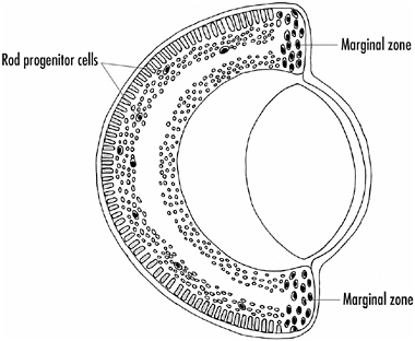
FIGURE 5-1A sketch of a fish eye showing the marginal zone where there are stem cells that divide and generate new neural and glial cells throughout the life of the animal. There are also some stem cells, called rod progenitor cells (arrows), found throughout the outer retina. These cells ordinarily generate new rod photoreceptor cells, but if the retina is damaged, they generate all retinal cell types, thus repairing the damage.
large lesions are completely repaired and vision is restored to that part of the eye.
Why the cold-blooded nervous system can regenerate itself and the mammalian brain cannot, or can only to a very limited extent, is not understood, but it is obviously an area needing further study. It is curious that the brains of cold-blooded vertebrates are much more plastic in terms of neuronal replacement and repair than mammalian brains, but at the same time they appear much more hard-wired and perceptually less plastic than mammalian brains. Recall the example of inverting prisms: Humans rapidly adjust to an upside-down world, but when a frog’s world is turned upside down, it remains that way. Is there a trade-off of one type of plasticity for the other? No one knows.
Cold-blooded vertebrate neurons differ from mammalian neurons in other ways, particularly with regard to the regeneration of axons. Whereas the axons of peripheral nervous system neurons
(that is, axons outside the spinal cord and brain) in both mammalian and nonmammalian species regrow after being severed, central nervous system axons in mammals do not. Thus, spinal cord injuries that cause the severing or loss of central nervous system axons result in a permanent paralysis of the body and limbs served by those axons. Not only motor function, but also sensation is lost. Such people can never walk again. If the spinal cord injury is just below the neck, they might also lose the use of their arms and require assistance breathing, necessitating the use of a respirator. The actor Christopher Reeve, whose spinal cord was crushed in a riding accident, is an example of this type of devastating injury.
It turns out that the determinant of whether an axon regenerates resides not in the axon itself, but in the glial cells that cover and contact the axons. All axons are ensheathed by glial cells, and the glial cells form a lipid layer (the myelin sheath) around most axons to insulate them. The glial cells in the peripheral nervous system that ensheath the axons are distinct from those in the central nervous system. If severed central nervous system axons are brought into contact with peripheral nervous system glial cells, at least some of the axons regenerate, as first shown by Albert Aguayo and his colleagues in Montreal. It seems that two things are going on. The peripheral nervous system glial cells appear to release substances that promote the regeneration of axons, whereas central nervous system glial cells release a factor or factors that inhibit the regeneration of axons. Identification of these factors is currently being attempted with some success, but the story is not yet complete.
In cold-blooded vertebrates, central nervous system axons do regenerate, so this is another avenue of research to pursue—to attempt to find differences in the nerve and glial cells between cold-blooded and mammalian species. Such studies might teach us not only why axons regenerate in one situation but not another but, even more importantly, how we might induce the generation of new neurons throughout the brains of warm-blooded animals.
How Plastic Is the Mammalian Brain?
Another issue is how much reorganization can occur in the mammalian brain, either young or old. I pointed out in the last chapter that when both the two hippocampi and their associated areas are destroyed, a person can no longer consolidate memories. Such people must live in the present and can never again remember new facts or events. There is no recovery.
Adults who have a massive stroke on the left side of the brain are often left mute and show little or no improvement in speaking abilities over time. However, a child can be very different. Occasionally, and fortunately it is very occasionally, it is necessary to remove virtually a whole cortical hemisphere in a child because of a condition known as Rasmussen’s encephalitis. This disease causes a debilitation of one hemisphere that results in severe disruption of the other. Surgical removal of the diseased hemisphere permits the other hemisphere to function. The question is how well these children do after surgery, especially if it is the left hemisphere with its language centers that is removed.
The answer is that they do remarkably well. They learn to speak again over months, and motor function on the right side also returns. Neither speaking itself nor motor function ever becomes entirely normal, but the amount of recovery is remarkable nevertheless. Language comprehension is better than speaking for such children and, as is expected, the younger the child is at the time of the surgery, the better and quicker is the recovery. No one older than 15 has ever regained much function.
In these children, one hemisphere takes on the work of both; a new hemisphere does not develop and fMRI scans show that the remaining hemisphere does not increase greatly in size and fill up the skull. Rather there is a large void where the removed tissue was, as shown in Figure 5-2.
Exactly what is going on in the remaining hemisphere is not known, but presumably there is extensive remodeling of the neural circuitry, growth of new branches by the neurons, and new synapse formation. This has not yet been explored.
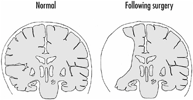
FIGURE 5-2On the right is a sketch of the brain after surgery of a young boy who suffered from Rasmussen’s encephalitis. Although much of the left hemisphere, including the language areas, was removed, the youngster regained speech and other functions normally controlled by the left hemisphere.
Is there a similar takeover of function by one part of the brain for another in all circumstances in children? The answer appears to be no. If, for example, there is a bilateral lesion of the cortex so that comparable areas of both hemispheres are damaged or destroyed, the functions mediated by those cortical areas do not recover. This has happened occasionally during operations on infants when oxygen levels in parts of the brain fell precipitously. The children were then left permanently mute and/or blind or with some other serious neurological deficit. It appears that recovery depends on a brain area in one hemisphere remaining intact.
Genes and Behavior
No one would deny that genes are critical for behavior, but the question of how and how much is a vexing one. It is almost needless to say that the relationship is an exceptionally complex one, and because of the enormous stakes involved—ranging from how we raise our children to how we deal with societal issues—the debate is hot and heavy. We read every day about a newly discovered gene that accounts for this behavior or that—from bed wet-
ting to homosexuality to doing mathematics. We also read reports from the other side of the debate, arguing that behavior is, as the title of one book indicates, Not in Our Genes. The truth is some-where in the middle, of course, and we are still a long way from sorting it all out.
One classic example often cited as showing a relation between genes and behavior is schizophrenia, a devastating cognitive disorder that results in thought and mood disorders, delusions and hallucinations, and the withdrawal from social interactions. The judgment of schizophrenics is markedly impaired and they can lose all contact with reality. It has been known for some time that there is an inheritance factor in susceptibility to schizophrenia, and the question is, how large is the inheritance factor? Studies of identical twins, whose genetics are the same, concluded that if your identical twin is schizophrenic, the chances that you will become schizophrenic are about 50 percent. On the other hand, fraternal twins have an incidence of about 15 percent and schizophrenia in the general population is only 1-2 percent.
“Ah,” says one side, “see how important genetics is for this behavioral disorder!” But the other side comes back with: “What about the 50 percent that don’t become schizophrenic? The genes are the same; environment is critical!” And, of course, both sides are right and so we are in a muddle. What can we say?
Huntington’s Disease—A Clearer Case
An inherited neurological disorder, Huntington’s disease, is a clearer example of how one gene can cause a devastating disease affecting behavior. The disease is fortunately rare—affecting about 1 in 10,000 people. It begins usually in young to middle-aged adults with the appearance of small, uncontrolled movements. The symptoms gradually increase in severity until the patient is confined to bed. Decreased mental capability, including memory loss and personality change, also occur over time, and death ensues 15-20 years after onset. Thus, the disease affects both motor control and cognitive processes.
The disease is inherited in a dominant fashion; that is, someone who inherits one copy of the gene is affected and everyone who inherits the defective gene comes down with the disease. This means that if one parent has the disease, there is a 50 percent chance that each offspring will be affected. And if one identical twin has the disease, the other is certain to get it (although such cases have not been reported, as far as I am aware).
In the 1980s, James Gusella and his colleagues at the Massachusetts General Hospital localized the gene for Huntington’s disease to human chromosome 4. It took them and others another 10 years to isolate, clone, and sequence the gene, illustrating that identifying and analyzing mutated genes is not a simple task.
The gene codes for a large protein, which was given the name Huntingtin, but its function still remains unknown. The protein is found in a variety of cells in the brain and elsewhere, but why it seems to particularly affect some neurons and not others, and some neural processes and not others, is very much a mystery.
The nature of the defective gene and protein has now been unraveled. The mutant protein has an excessive number of a particular kind of amino acid near one end of the molecule. The amino acid is called glutamine and it is coded in the gene’s DNA by the nucleotide sequence cytosine-adenine-guanosine (CAG). Thus in the defective gene, the CAG sequence is repeated over and over again. In normal people, there are some—between 6 and 34—CAG repeats in the gene. However, people with Huntington’s disease have from 37 to more than 250 CAG repeats. There is, however, a bit of variation here. Some people with Huntington’s disease have just 36 repeats and a few older people who have 39 repeats appear quite normal. But anyone with more than 40 repeats definitely gets the disease.
Observers have long recognized that there is considerable variability in the onset and progression of the disease and some—particularly those arguing that much more is involved in behavior phenomena than single genes—suggest that the variations are due to other genes or to environmental effects. Variation in the effects of a single mutated gene is often termed gene penetrance. Recent
data suggest that in Huntington’s disease the variability relates mainly to the structure of the defective gene.
This became clear when it was noticed that the onset, progression, and severity of the disease increased in some families over generations, especially when the disease was passed on from the father. When the defective gene was looked at in different generations of these families, the number of CAG repeats increased with succeeding generations. It was then possible to relate the severity of the disease to the number of CAG repeats, and this finding appears to explain much of the variability in the disease. The more CAG repeats in the gene—and hence the more glutamines in the protein—the earlier the onset and the faster the progression of the disease. Why the number of CAG repeats increases in succeeding generations of these families is not understood.
Several neurodegenerative diseases have now been linked to increased numbers of CAG repeats in specific genes but we don’t know why this causes problems. It might be that proteins with excess numbers of glutamine molecules accumulate in the cells—the cells have difficulty in breaking down the excess and abnormal protein—and become toxic to particular neurons. Indeed, there is evidence that consecutive and excessive glutamines in a protein make it “sticky,” so that one possibility is that the mutant proteins stick to each other and or to other proteins, causing an accumulation of protein. Another possibility is that fragments that result from breakdown of the mutant protein are toxic. Thus, there might be a gradual accumulation of toxic protein aggregates in certain cells, or toxic products might accumulate during protein breakdown, and both situations could lead eventually to the death of the neuron.
Complex and Cognitive Behaviors
I know of no non-disease-related behavioral or cognitive trait in humans that can yet be related to a single gene. It is generally believed that complex behavioral traits result from the products
of many genes. At first glance, a promising exception is FOXP-2, the gene that causes the speech and language disorder in the large human family described in Chapter 3. The gene defect is found in every affected family member, but not in any normal family member nor, so far, in any unrelated normal person. One unrelated person who has the disorder has also been found to have the same gene defect. As noted in Chapter 3, the gene is dominantly inherited and leads to defects in speech and language, including grammatical skills.
The function of the gene is not known, although it has been proposed that it is important in the development of circuitry related to speech and language skills. Broca’s area, for example, could be one possibility for the site of action of the gene, although there is no evidence for this. The important point is that, whereas this is a single gene, it codes for a transcription factor, and transcription factors can turn on and off many genes. Thus, it is likely to be not just a single gene and protein involved here, but ultimately many genes and many proteins probably playing a role.
Genes, Environment, Development, and Behavior
So far, the examples I have chosen stress the importance of genes in behaviors, but this is only one side of the story. What can we say about the other side, environment and other developmental factors on behavior?
My current research is mainly with zebrafish, small, freshwater, bony fish that are convenient for genetic studies. Zebrafish are vertebrates—animals with backbones—as are we, and they are at present the only vertebrate with which we can do certain kinds of genetic studies. We can inbreed zebrafish so that they become virtual clones. In other words, the genetics of the fish we use are essentially identical.
My laboratory is interested primarily in the visual system, especially the retina. We produce mutations in the fish’s genome by altering the genetic material in the fish with specific chemicals and then we look for changes in the structure, function, and
development of the retina. We find single-gene mutations that affect the retina fairly frequently; many of these alter the visual behavior of the animals, not unexpectedly. Disrupting some aspect of the function of the light-sensitive cells in the retina, the photoreceptors, or other retinal neurons, obviously decreases the animal’s ability to see properly, and this affects visual behaviors. For example, we have found single gene mutations that render animals completely blind, partially blind, color-blind, or movement insensitive. In many cases we have identified the genetic nature of the defect, but again this is not at all surprising.
However, we have also made a number of observations that bear on the question of genes, environment, and other possible factors on higher-order behaviors. Let me describe one inadvertent experiment carried out by a young colleague. He was interested in observing zebrafish in a tank and began with 12 young adult fish, all of which came from the same cross. That is, they were all siblings, from the same parents, of the same age and raised under identical conditions. Genetically they were as similar as zebrafish can be and they were raised under as identical conditions as is possible—in the same tank, at the same temperature, and so on.
To make his observation tank more interesting, my colleague put an elongated plant at one end. Shortly thereafter, as he was observing the fish, he noticed that one fish was swimming in the open, whereas the other 11 were staying in or close to the plant. Curious, he disturbed the surface of the water in the tank, which usually indicates to the fish that food is coming. The fish charged out from the plant and milled around looking for food. There was none, but then my colleague noted a most interesting phenomenon. The fish that was originally swimming in the open began to chase the others back into the plant. This took a few minutes, but he or she eventually succeeded. It was clear that this fish was running the show!
We speak of this behavior as dominance and it is commonly observed in fish and many other animals. The interest here is the fact that all the fish were essentially identical genetically—not
100 percent identical, but close to that. Genetic factors presumably can be ruled out as playing a role here. We might suppose that it is an environmental effect, but here, too, we are stumped. The animals were all raised identically. There were some size differences among them, but that didn’t seem to be key in follow-up experiments as to who would be the dominant fish. The answer is still not clear, but this example makes the point that either extremely small genetic differences or unrecognizable environmental differences can make a big difference in an animal’s behavior.
But possibilities other than genetics or environment might also be at play here. Chance, for example, could play a significant role. That is, during brain development many things go on—cell specification and differentiation, cell migration, axonal pathfinding, pruning of neurons, rearrangement of synapses, and so forth (Chapters 1 and 2). Some chance variation in these processes in genetically identical animals raised under identical conditions is likely to occur and this could give rise to significant behavioral differences. The bottom line is that it can be very difficult to cull out behavioral, genetic, or other (developmental) differences among organisms and to assign these differences to specific behavioral traits.
Another set of factors that might be involved here are the so-called “epigenetic” factors that regulate how genes are expressed in cells and how much protein is made. We know, for example, that gene expression can be modulated by enzymes that add methyl groups onto the DNA bases, or how the DNA molecule is made accessible for the expression of its genes. In the nucleus, the long DNA molecules are wrapped tightly around specific proteins to form a highly condensed structure called chromatin. There are enzymes, chromatin-remodeling complexes, that unwrap portions of DNA from chromatin to make it accessible to transcription and other factors that lead to expression of the genes on that bit of DNA. We still know little about these epigenetic factors, but obviously variations in them can alter gene expression quite significantly. In other words, it is not simply gene sequence—that is, the particular protein produced from a specific gene sequence—that
is important, but how much protein product is produced from a specific gene, and this amount can be influenced by epigenetic factors.
Twin Studies
What has long been considered the gold standard for uncovering the relative influences of genes and environment on behavior in humans is the study of identical twins reared apart. However, this field has been fraught with controversy, including claims of scientific fraud against one of its pioneers, the Englishman Cyril Burt, who studied identical twins reared apart from the 1920s to the 1960s. And at least some of the claims of fraud against Burt appear to be true. However, over the past 20 years, the field has been revisited by Thomas Bouchard and his colleagues at the University of Minnesota, and their work is careful, detailed, and compelling. It is based on a study of more than 100 sets of reared-apart twins or triplets, mainly from the United States and the United Kingdom, but twins from six other countries were examined as well.
The twins were brought to Minneapolis where they were put through 50 hours of medical and psychological testing over the course of a week. In some cases the twins had had contact with one another over the years, but in others they did not. In some cases, the twins were raised in families with quite similar back-grounds and values, but in others they were not. Regardless, the main finding of the study was that identical twins raised apart are about as similar as identical twins reared together. The conclusion drawn was that being reared in a different environment does not appear to influence many behavioral traits and therefore genetic factors must play a significant role in determining a variety of behaviors.
This is not to say that environmental or other factors do not play a key role in determining human behaviors. For no trait examined was the result identical among the pairs of identical twins, whether they were raised together or apart, and the correlations
ranged from low (0.34) to high (0.78). (These numbers mean that as little as 11 percent to as much as 61 percent of the variability is genetic—the correlation is squared to arrive at the variance.) Although it is difficult to put hard numbers on all of this, the conclusion drawn is that about 50 percent of the variation in personality traits is genetic; the other 50 percent is due to environmental or other factors. It should be noted, however, that many psychologists believe an heritability estimate of 50 percent for personality traits in humans is too high, based primarily on animal (rat) studies in which heritability of behavioral traits has been shown to be much lower (<0.2).
The big surprise at first glance from the twins study was that the home environment did not have more effect on behavioral traits. There are two comments to be made here. First, the rearing environments were probably not all that different among the cases of reared-apart twins. We raise children in much the same way throughout the developed world, and all the twins came from the developed world. It is when a rearing environment is deficient and children are deprived that we see substantial differences in behaviors that can be permanent.
The Romanian orphans are a case in point. As a result of harsh laws banning birth control and abortion in Romania in the 1970s, more than 100,000 unwanted children were born but then abandoned in orphanages. They were given little environmental stimulation and had little personal interaction with caring adults. After the fall of the Ceausescu regime, many of these children were rescued and placed in adoptive homes in the West. Those adopted at younger ages (before six months) have done reasonably well, but children adopted at older ages (one to three years) have shown persistent personality defects into their teenage years. The guess is they will never recover emotionally or socially as a result of that early deprivation.
The Romanian orphan situation is reminiscent of the experiments of Harry Harlow of the University of Wisconsin in the 1950s and 1960s. Working with infant rhesus monkeys, he showed that these monkeys often demonstrate lasting adverse effects if
deprived of social and maternal interactions early in life. (Other monkey species, on the other hand, are less affected by social or maternal isolation.) Rhesus monkeys show depression and other behavioral disturbances when isolated from their mothers and other monkeys at an early age, and often these are permanent effects, although if deprived monkeys are subsequently placed with normal infant monkeys, they do appear to recover a great deal of normal behavior. In some experiments, the infant monkeys were housed in close proximity to other monkeys, both young and old, so that they could see, hear, and even smell them. Nevertheless, the monkeys deprived of contact with other animals showed severe behavioral disturbances. Physical interaction appears to be important in the nurturing of many young primates and other mammals. Harlow showed, for example, that maternally deprived infant monkeys would cling to an artificial terry-cloth mother and ignore a wire-frame mother, even if the wire-frame mother was the source of food—its milk bottle was attached to the wire-frame mother.
Of interest in this regard is the recent finding that mouse pups that lack a receptor protein activated by opiates show much less distress when isolated from their mothers as compared to normal pups. This suggests that during bonding between mother and infant, endogenous opiate-like substances are released in the infant’s brain, activating the receptors and leading to pleasurable feelings that contribute to the bonding between mother and child. Opiates are well known to alleviate pain as well as provide pleasurable feelings, and thus the lack of activation of opiate receptors in the brains of socially isolated animals could, perhaps, result in painful feelings that contribute to the distress shown by such infants.
Other experiments with rats have shown that newborn animals that receive no touch stimuli during an early critical period show increased levels of the stress hormone cortisol in response to various challenging situations that the animal encounters even when an adult. Excessive levels of cortisol have also been found in the Romanian orphans when they were subjected to a some-
what stressful event—being examined by a doctor unknown to them, for example. As was noted earlier in the chapter, increased levels of the stress hormone leads to a decreased proliferation of neurons in the hippocampus, and there is also evidence that increased levels of the stress hormone cortisol cause brain damage in animals, including neuronal cell death. In aging humans, increased cortisol levels have been linked to difficulties in performing certain memory tasks. (Chapter 6 discusses this further.)
The second comment with regard to the apparent lack of effect of home environment is that children themselves play a significant role in the type of environment they experience growing up. They select the parent to whom they respond more closely, their friends, and the activities they most enjoy and prefer to do. Children demonstrate different temperaments, and parents usually respond predictably to these temperaments. One doesn’t hug excessively a child who obviously dislikes hugging. I am not saying, and others have made this point in much more detail and more persuasively than I do here, that the home environment is not important; it certainly is, as the experience of the Romanian orphans so clearly demonstrates. However, as long as the home environment is supportive and caring, genetic factors play an important role in the determination of behavioral traits.
IQ and Genetics
Of all the topics touched upon in this book, perhaps none is more contentious than IQ and inheritance. The topic generates much more heat than light. Nevertheless, it is one that cannot be entirely ducked in a book that deals with nature-nurture issues.
The first question that might be raised is whether the concept of IQ has validity. Can intelligence be realistically and reliably measured and given a number, usually termed “g”? Everyone agrees that many cognitive skills are not included in present IQ tests, so what does an IQ score really mean? There is no question that IQ does predict such things as success in school, job performance, and even future economic and social status, but, and this
is a very big “but,” not perfectly. And it is the big “but” in the last sentence that is the hang-up—how well does IQ predict these things, and is too much left out in determining a single “g” number for someone?
A number of psychologists, especially Howard Gardner of Harvard University, espouse the idea of multiple intelligences, and I find this notion attractive. When I compare my wife’s intellectual abilities to my own, I am struck by her obvious superior abilities in some areas compared to mine, and I believe I might have some abilities superior to hers (although I dare not tell her so). Gardner has proposed seven categories of intelligence: linguistic, musical, logical-mathematical, spatial, bodily-kinesthetic, intrapersonal, and interpersonal. One of the strengths of this categorization—that gives it some neurobiological meaning—is that each form can be selectively affected by one or another type of brain damage, as Gardner points out. Others have proposed somewhat different categories, but the idea is basically the same—that we all differ behaviorally and cognitively in different ways and this should be taken into account in evaluating intelligence.
However, at the moment we are stuck with one IQ score and the question is how much of IQ can be attributed to heredity, to environment, or to other factors. Needless to say, these are complex relationships that are not easily teased apart. Bouchard’s twin studies indicate that the variance in IQ due to genetic variation is high—60-70 percent. Earlier studies had suggested somewhat lower values—40-50 percent—with regard to the heritability of IQ, but Bouchard’s results were obtained from middle-aged adults, and recent findings suggest that the correlation of IQ and inheritance increases with age. Indeed, there is good evidence that IQ is more environmentally linked in children, youngsters, and young adults than in middle-aged or older people. For example, IQ scores in children correlate with the education of their parents in every society studied.
One can also correlate lower IQ levels with lower birth weights. Alcohol and drug use as well as cigarette smoking during pregnancy also seem to decrease IQ levels. Lead exposure both in
pregnancy and in young children also leads to lower IQs, as does a deprived environment during the early years. It is the deprived-environment children who seem to benefit most from social programs such as Head Start and whose IQs appear to be raised significantly by the programs. But here again, the data are somewhat murky and often disputed. The effects of programs such as Head Start seem to peter out as children get into school. Schooling itself appears to influence IQ; each year of schooling might increase IQ by two to four points.
The bottom line here is that it is the most deprived children whose IQs can be raised most by environmental intervention. The IQs of children raised in solidly supportive and caring environments are less environmentally related. But exactly how much the “less” is is hotly debated, and neurobiology can contribute nothing to the debate.
I end with a most curious observation with regard to IQ scores that needs, in my view, much more examination. And this is the fact that IQ scores are gradually rising—at a rate of about three points per decade as shown in Figure 5-3.
What might this mean? Are we becoming more intelligent or just better at taking the tests—training our children to deal better with them. The rise of IQ scores over time was summarized in the mid-1980s by James Flynn in New Zealand and is often known as the Flynn effect. The increase is substantial: In 1932, about 2.5 percent of the population scored above 130 on the IQ test and were categorized as in the “very superior” group. By 1997, about 25 percent of the population would score at this level if they were to take the 1932 test. Has intelligence risen by that much? How could that be possible?
Both sides of the issue are argued. Some suggest that we are smarter because we are bigger and that means a bigger brain. In the industrialized world, we have been gaining an average of 1 cm in height per decade, thought to be due to better health and nutrition. The implication here is that we are smarter than our parents and our children are smarter than we are. Some are comfortable with this notion, but others are not.

FIGURE 5-3The Flynn effect: There has been a consistent increase in mean performance on a variety of IQ tests from the early 1930s to the present. Why this is so is not understood.
On the other side are those who insist that it is an environmental effect—schooling and training children to take the tests is one possible answer, but has schooling changed enough since the 1930s to make this much of a difference? Schooling relates more to content than reasoning and it is in reasoning that the test results show the greatest increases.
An interesting factor that has been proposed and is clearly worth exploring further is the visual environment to which our children are now exposed compared to those we or our parents experienced. Clearly, the visual environment available to us is more complex and has become increasingly more so since the 1920s when movies were introduced. Television was another milestone enhancing the richness of our visual world, and then computers and computer games arrived in the 1980s. Of course, the computer introduces more than an enriched visual environment, so it will be interesting to see if IQ scores tend to rise slowly over the next few decades or, indeed, rise even faster. There might be neurobiological implications here if it becomes possible to determine the effects of such enriched environments on brain structure and/or function.
An intriguing study recently published in the journal Nature bears on this issue. When subjects between the ages of 18 and 23 who played action video games for at least one hour per day four days per week for the previous six months were tested on visual attention tasks, they performed significantly better than subjects the same age who did not play such games. For example, the video-game players were superior in determining how many objects were present in a briefly flashed display. The differences in performance by the video-game-playing group compared to the nonplaying group were highly significant. Improvement in such tasks by the nonplaying group was achieved by having them play a specific game for one hour per day for 10 consecutive days. These findings convincingly demonstrate the effects of training on certain cognitive tasks.