Understanding and Preventing Violence, Volume 2: Biobehavioral Influences (1994)
Chapter: Afferent Connections
establishes the anatomic substrate by which jaw opening and jaw closing are controlled by the perifornical hypothalamus for the biting component of the attack response. Furthermore, the projection to the locus coeruleus provides the possible substrate by which noradrenergic pools from this nucleus can be activated to generate the arousal component of the attack response. Other research (Shaikh et al., 1987) suggests that the predatory attack system at the level of the PAG (see Figure 4) is organized along several possible plans: (1) that the PAG serves as a feedback system to the perifornical hypothalamus from which the primary integrated output to the lower brain stem is organized; and/or (2) that the PAG serves as a feed-forward relay of the perifornical hypothalamus, but does so through the use of short axons passing to the tegmental fields. The precise function of such fibers remains unknown, but they might comprise part of a multisynaptic pathway (presumably via reticulospinal fibers) for the regulation of autonomic and somatic motor components of the predatory attack response. The projection to the raphe nucleus, however, is more likely to be associated with the modulation of the attack response. This judgment is based on the fact that stimulation of the raphe nucleus has been shown to suppress predatory attack (Shaikh et al., 1984) and that administration of para-chlorophenylalanine (a monamine suppressor) can facilitate the occurrence of this response (MacDonnell et al., 1971).
In this discussion, the likely anatomic substrates are described over which the autonomic and several of the somatic motor components of the predatory attack response are expressed. However, little has been said of the anatomic substrates governing how the hypothalamus might regulate visual processes central to the organization of the attack response. Research by Pott and MacDonnell (1986) and by Ogren and Hendrickson (1976) provides suggestions as to how this may be accomplished. The projection from the posterior lateral hypothalamus to the pulvinar represents the initial limb in a disynaptic pathway from the hypothalamus to the visual cortex that may be essential for the integration of visual information as well as for controlling visual pursuit movements during the attack sequence.
AFFECTIVE DEFENSE BEHAVIOR
Afferent Connections
As noted earlier in this chapter, the sites in the brain at which affective defense reactions can be elicited have been identified
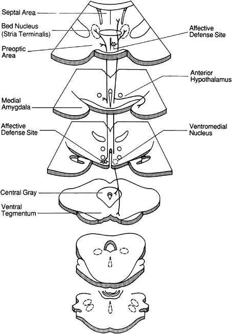
FIGURE 4 Diagram indicating the principal ascending and descending projections of the medial hypothalamus associated with affective defense behavior. Of significance is the fact that fibers mediating this response arise from the ventromedial nucleus and primarily project rostrally into the anteromedial hypothalamus, medial preoptic region, and bed nucleus of the stria terminalis. Note that the fibers that supply the midbrain periaqueductal gray in association with affective defense arise primarily from the anteromedial hypothalamus.
SOURCE: Siegel and Pott (1988).

